Precision Surgery with Advanced Robotics
Minimally invasive robotic surgery for faster recovery and better outcomes.
Riverside is committed to providing the best healthcare possible to each and every patient we encounter. This commitment means a dedication to technology and the application of robotics in a surgical setting. The benefits from robotic surgery have shown to result in faster recovery time, smaller incision size, and less blood loss.
Riverside has been at the forefront of robotic-assisted surgery since introducing its first da Vinci surgical system more than 15 years ago.
Riverside utilizes a number of robots in surgical settings.
Robotics at Riverside
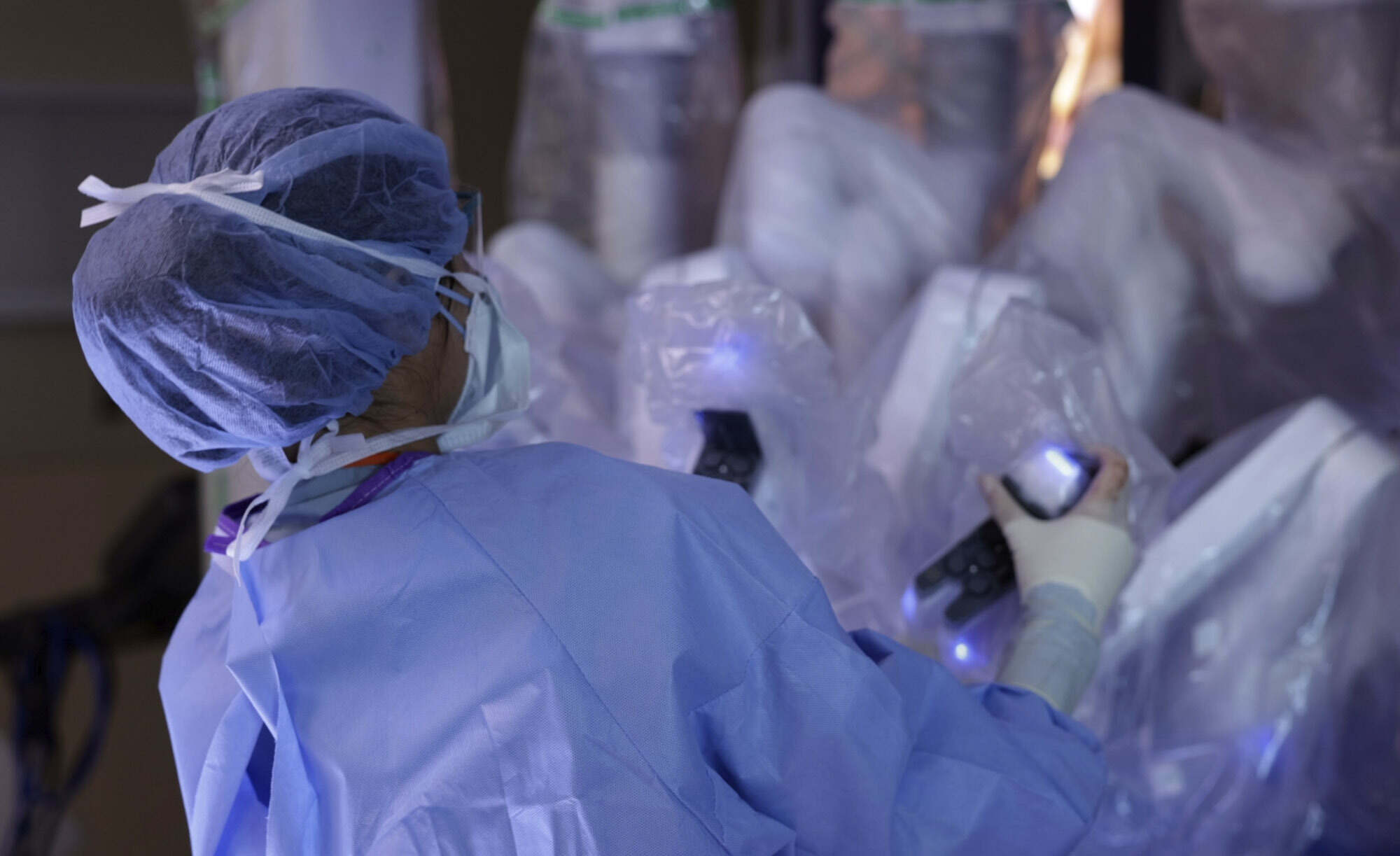
Robotic surgery, or robot-assisted surgery, allows doctors to perform many types of complex procedures with more precision, flexibility, and control than is possible with conventional techniques.
Watch the Video|
|
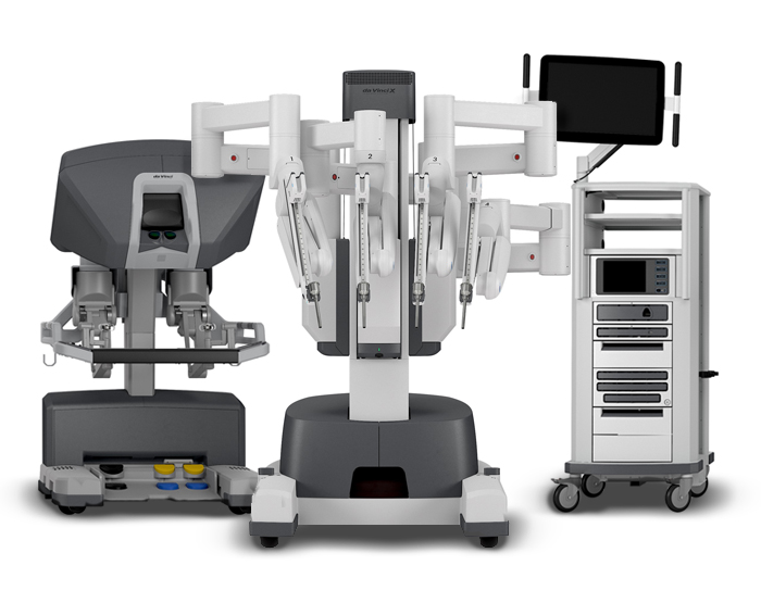
DaVinci XIUsed by General Surgery, Urology, OB-GYN The newer DaVinci XI system features the addition of a fourth arm to the system. Along with this, all of the arms are mounted onto an overhead boom that can rotate and pivot into virtually any position. The arms can even be disconnected and reconnected mid-procedure if the doctors feel like swapping them around. The robotic system acts as an extension of the surgeon's eyes and hands, giving the physician 3-D magnified vision and 360° dexterity of four arms, which allows for more effective, precise surgical movements. The surgeon is 100% in control of the robot, which translates their hand movements into smaller, more precise movements of tiny instruments inside the patient’s body. Additionally, the immersive 3D-HD vision system provides surgeons a highly magnified view, virtually extending their eyes and hands into the patient. Surgeons specifically trained on the DaVinci Xi at Riverside Medical Center are able to perform numerous kinds of abdominal, gynecological, and urological operations. This specifically means surgeons can offer robotic-assisted hernia repair, complex abdominal wall reconstruction, anti-reflux surgery, gallbladder surgery, surgery of the small and large intestine, weight-loss surgery, prostate surgery, kidney surgery, and surgery of the ovaries and uterus. |
|
|
|
|
|
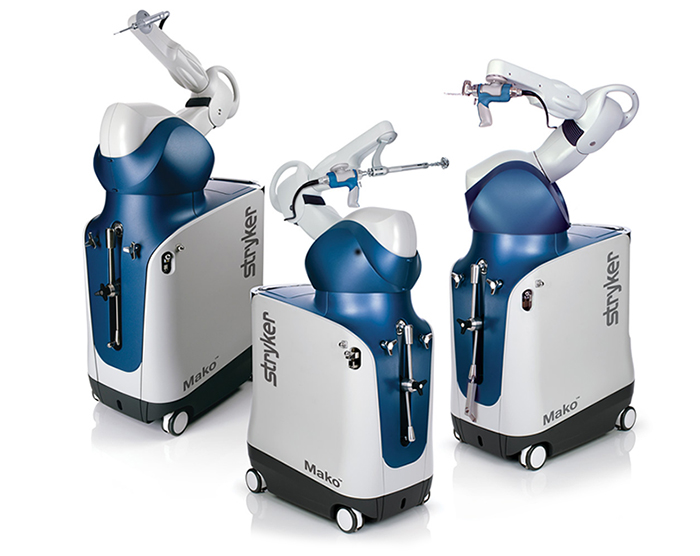
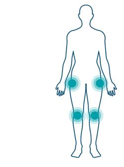
MakoUsed by Riverside Orthopedic Specialists Mako SmartRobotics is an innovative solution for many suffering from painful arthritis of the knee or hip. Mako uses 3D CT-based planning software so your surgeon can know more about your anatomy to create a personalized joint replacement surgical plan. This 3D model is used to preplan and assist the surgeon in performing the joint replacement procedure. In the operating room, the surgeon follows your personalized surgical plan while preparing the bone for the implant. The surgeon guides Mako’s robotic arm within the predefined area, and Mako’s AccuStop technology helps the surgeon stay within the planned boundaries that were defined when the personalized pre-operative plan was created. By guiding the doctor during surgery, Mako’s AccuStop technology allows the surgeon to cut less by cutting precisely what’s planned to help protect healthy bone. It’s important to understand that the surgery is performed by an orthopedic surgeon, who guides Mako’s robotic arm during the surgery to position the implant in the knee and hip joints. Mako SmartRobotics does not perform surgery, make decisions on its own or move without the surgeon guiding it. Mako SmartRobotics also allows your surgeon to make adjustments to your plan during surgery as needed. |
|
|
|
 |
|
|
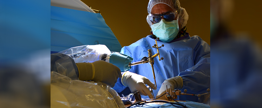
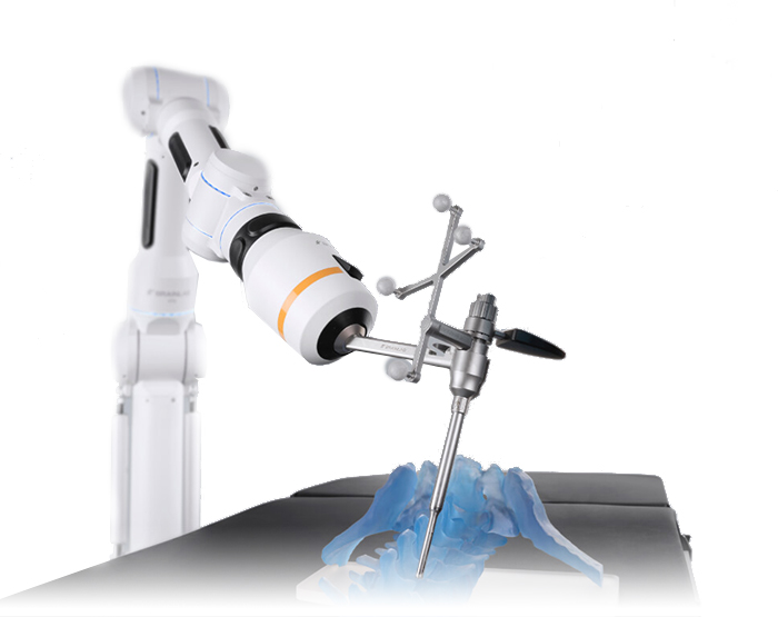
CirqUsed by Riverside Brain & Spine Institute The Cirq, dubbed “Cirqules” after the Roman god known for strength, is an essential part of the team of leading-edge technologies Riverside Healthcare utilizes to deliver expert patient care. Inspired by the form of the human arm, Cirq is a reliable and intuitive assistant during surgical procedures. With robotic technologies Riverside's Neurosciences Specialists can focus on the delicate skillful parts of the procedure, while the Cirq becomes like a third arm, aiding in accuracy and placements. During an operation, the surgeon moves and locks the robot into place to guide and stabilize surgical steps. By having an extra, software-driven hand, the surgeon can focus on the procedure with both human hands. |
|
|
|
 |
|
|
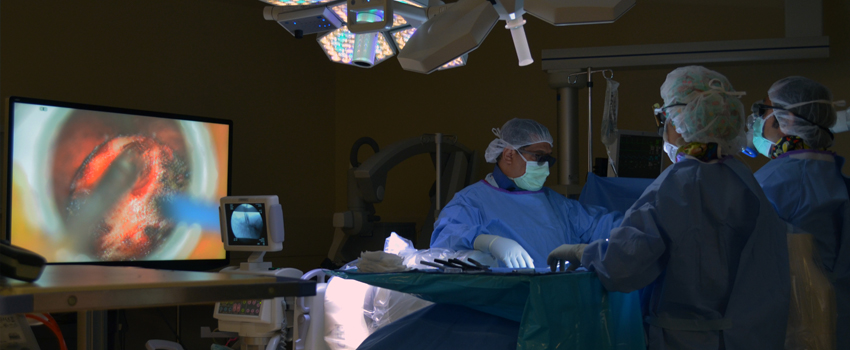
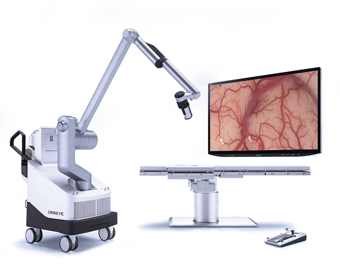
ORBEYEUsed by Riverside Brain & Spine Institute The precise 4K-3D images from the ORBEYE surgical microscope provide 26x magnification imaging to aid in life-saving procedures, all displayed on a large 55-inch 4K-3D monitor for the entire surgical team to view in real-time. The enhanced imaging provides high-resolution details of structural and surrounding tissue, tumors, blood vessels, and other features, allowing the team to tailor appropriate surgical interventions. This all happens through dime-sized incisions, which has a major impact on the recovery time for the patient. Additionally, the ORBEYE has the potential to reduce surgeon fatigue by eliminating the need for extensive microscope eyepiece viewing and allows for a more comfortable, upright working posture. The ORBEYE microscope unit itself is much smaller, too, allowing for additional operative space. The ORBEYE 4K-3D Microscope, developed in a joint venture between Olympus Corporation and Sony Imaging Products & Solutions, Inc., is used for skull-based and spine procedures and enhances minimally invasive techniques used by neurosurgeons in some of the most complex and lengthy neurosurgeries. |
|
|
|
 |
Augmented Reality with Zeiss Kinevo MicroscopeUsed by Riverside Brain & Spine Institute Riverside has partnered with Brainlab to bring this technology to the region by using the company's Zeiss Kinevo Microscope. This new technology provides Riverside's Neurosciences Specialists with a projected image of a patient's internal structures, allowing surgeons to gauge where they are in the procedure and focus directly on an area. This technology gives surgeons clearer visuals by compiling previous scans as well as real-time data. Riverside surgeons can choose to use the augmented reality either by a headset or nearby screen. The fast image processing system results in real-time visualization with no delay between the surgeon’s movements and the screen’s display. This allows for smooth viewing and precise instrument placement for the operation of a specific location. |

